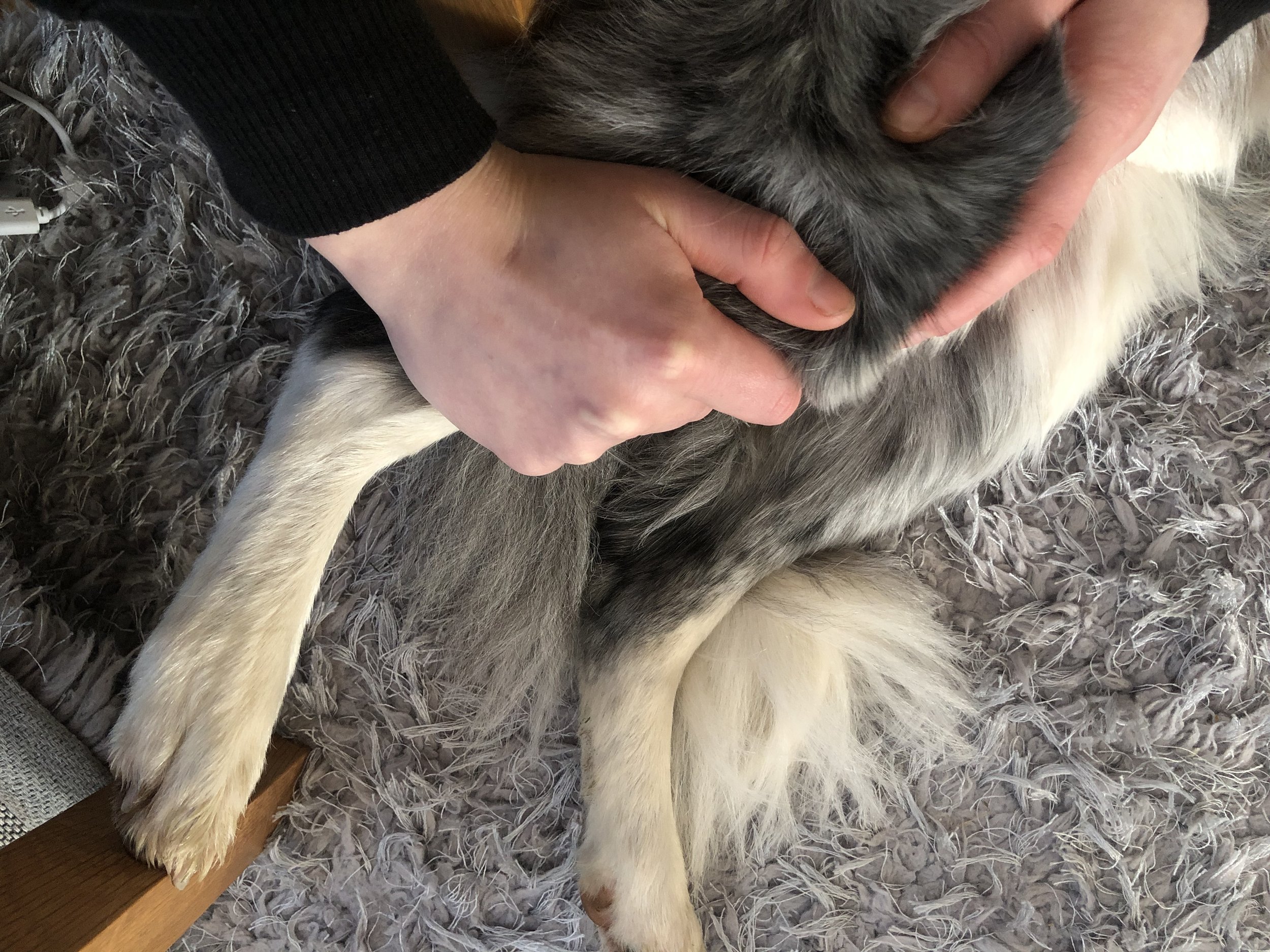
Canine Cruciate Ligament Damage
The Cruciate Ligaments are two bands of tough fibrous tissue located within the stifle (knee) joint. They join the femur (thigh bones) and tibia (below the knee bone) to allow the joint to work as a stable hinge joint. They are arranged as a cross, preventing the tibia from slipping forward in relation to the femur and preventing overextension and rotation. The cranial cruciate ligament runs from the inside to the outside of the knee, and the caudal cruciate runs from the outside to the inside.
Due to the joints movement being restricted by ligaments instead of interlocking bone, this makes it vulnerable to trauma and degeneration. The most common cruciate ligament damaged in the dog is the cranial cruciate ligament (CrCL). If damaged the knee becomes wobbly and very painful.
The CrCL is often injured through twisting, skidding, jumping, and turning awkwardly. Rupturing the CrCL is extremely painful. More commonly, the ligament fibres weaken over time and rupture occurs due to long term degeneration. This is often seen in larger breed dogs. Obesity is also a predisposing factor, as well as dogs with other knee problems, such as arthritis or patella luxation. Dogs who rupture one CrCL are likely to rupture the ligament in the other knee.
When the CrCL loses its normal mechanical function, pain and lameness start. When weight is taken onto the limb, the femur will roll down the tibial slope, causing pain and an inability to use the knee normally. If the slope is naturally steep, often seen in straight legged dogs, then this can be a predisposing factor as well.
If your dog traumatically ruptures their CrCL you can expect to see them suddenly stop while running, possibly cry out in pain, and be unable to fully weight bear on the affected limb. If damaged through fibre deterioration, some of the first signs you are likely to notice include; limping (especially during or following exercise), stiffness when getting up, back leg pain, swelling around the knee and an unusual walking gait. If you notice your dog is limping you should always visit your vet or contact your veterinary physiotherapist.
Diagnosis
If CrCL damage is suspected, your vet will perform a hand-on assessment of your dog's knee, which will include a specialised test called the cranial drawer which assesses abnormal forward movement of the tibia bone (lower leg bone) in front of the femur (thigh bone). An x-ray may also be necessary, and an exploratory arthroscopy (keyhole surgery) may be used to confirm the diagnosis, and look for possible cartilage tears and other problems.
Treatment
Most dogs will eventually require surgery to correct this painful injury and restabilise the knee. In some dogs weighing less than 10kg the ligament may heal by itself following 6 weeks of strict crate rest and an intensive conservative management programme.
Conservative management is only considered for small dogs and patients that are not appropriate for surgery, e.g. with heart disease, immune conditions etc. Management is often focused around weight management, physiotherapy, exercise modification and medication (anti-inflammatory painkillers). The same techniques are important in short-term management before surgery and during rehabilitation after surgery.
Surgery
There are various surgical techniques used to stabilise the knee joint. All will involve an inspection of the joint and removal of ruptured ligament fragments and repairing of meniscal cartilage if necessary. The most common surgical solutions are a tibial plateau levelling osteotomy (TPLO) and tibial tuberosity advancement (TTA).
Ligament replacement techniques, commonly used in humans, are not as commonly used in dogs because they have a poor chance of repair. This is because the replacement tissues are not as robust as the original ones, and the unfavourable position that caused the ligament to rupture in the first place is not corrected.
Newer surgeries render the CrCL redundant by changing the geometry of the knee joint so it's no longer needed to maintain stability. These surgeries are especially beneficial for larger dogs and have a high success rate.
TPLO thrust from Fitzpatrick Referrals on Vimeo:
This video illustrates what happens when a TPLO is perfomed.
During a TPLO first a radius cut will be performed in the top of the tibia bone. Then, the tibia bone is rotated so the slope is no longer present, achieving a level tibial plateau. The bone is then fixed in place using a bone plate and screws
A TTA surgery is similar,involving a cut in the tibia. However, its aim is to alter the direction of traction from the quadriceps muscle group, producing a force across the knee joint that neutralises the tendency for the femur to roll down the slope of the tibial plateau.
Because bone healing is more efficient than ligament healing, you can expect dogs to start weight bearing 1-3 days after a TPLO or TTA surgery. The success rate of these surgeries is over 90%. The risk of complication is low, but includes infection and mechanical failure due to too much exercise before the bones have healed properly (6 weeks).
Post operative care is very dog dependent but will likely include limited exercise for 6-8 weeks after surgery. Good function should ideally return within 3 months, if a rehabilitation plan is followed and reinjury does not occur. Arthritis is likely to develop as your dog ages (as with any surgery), which is worth bearing in mind and preparing for.
Preventing
Sometimes CrCL degeneration cannot be prevented, however, there are some predisposing factors that can be reduced by home management. These include, keeping your dog slim and exercising your dog sensibly (build exercise up gradually, keep consistent, limit strenuous exercise like jumping, skidding and chasing).


